
Abstract
Out-of-specification (OOS) results were reported by a contract lab in the in vitro adventitious agent assay (AVA) for two products manufactured using mouse myeloma cells in perfusion bioreactors. Cytopathic effect observed for test article-inoculated MRC-5 monolayers resembled foci seen in tissue culture cells infected with transforming viruses. All reasonable known technologies, including highly sensitive, state-of-the-art methodologies and multiple, redundant, and orthogonal methods, were deployed to screen broadly for potential viral and microbial contaminants. Due to the appearance of apparent foci, testing for murine, bovine, and human polyomavirus contamination was heavily represented in the analytical investigation. The results obtained in this extensive screening provided convincing evidence for the lack of an infectious viral or other biological agent. Although the initial investigation produced no reason to invalidate AVA yielding OOS results or to suspect an assay artifact, an extended evaluation revealed several irregularities at the contract test lab reporting the OOS results. The extended investigation also included attempts to reproduce OOS results at alternate contract testing labs and an inter-laboratory study in which methodological differences in the AVA at the three different contract labs were investigated. Only the contract lab initially reporting the OOS results reported foci during this extended evaluation. The results of the inter-laboratory study suggested that the foci artifact might be attributed to the prolonged exposure of the MRC-5 monolayer to cell debris present in the test article. Confocal immunofluorescence microscopy and transmission electron microscopy were subsequently used to provide convincing evidence that the foci observed in test article-inoculated AVA wells were composed of a core of degraded myeloma cell debris covered by one or more layers of MRC-5 cells. The observation that the foci were detected in the AVA at a contract lab where the MRC-5 monolayer is exposed to production cell line debris for a prolonged period strongly suggests that these foci form when MRC-5 grow over the cell debris present in the test article. The cumulative results of the investigation supported the conclusion that the OOS results were artifacts of the AVA test system and not a result of contamination with a virus or other biological agent. Testing was discontinued at the contract lab generating the OOS results and validated at a second contract lab. Manufacturing resumed in consultation with health authorities. The lots were retested following a standard operating procedure (SOP) already in place and ultimately dispositioned for use in normal distribution channels.
Introduction
In late 2008, out-of-specification (OOS) results were reported during viral safety release testing of samples derived from two manufacturing processes at Centocor. Both of the manufacturing processes used mouse myeloma production cell lines in perfusion bioreactors. One of the processes supported a late phase development product that used no animal-derived raw materials in the manufacturing process from cell banking through fill finish. The other produced a commercial product that included animal-derived raw materials in the production process.
OOS Results Obtained in an In Vitro Adventitious Agent Assay
The OOS results occurred in the in vitro adventitious viral agent assay (AVA). Health authorities require that sponsors evaluate biological products in an AVA by inoculating test article onto indicator cell lines that are susceptible to a wide range of relevant viruses, then subsequently observing the inoculated cultures for both cytopathic and hemadsorbing viruses. At the time of the OOS report, an AVA was conducted at Contract Lab A where the assay previously had been validated. The test article used for this AVA was a free-thaw lysate of production cells in their conditioned media derived from the final harvest of the perfusion bioreactor. Assay growth media was used as a negative control and parainfluenza virus 3 (PI-3) was included as a positive virus control. The test and control articles were inoculated into six well assay plates containing MRC-5, Vero, HeLa, and mouse myeloma indicator cells. Following 14 day incubation, the plates were evaluated for cytopathic effect (CPE), hemadsorption, and hemagglutination. See Figure 1 for an illustration of the assay.

In vitro adventitious agent assay (AVA) description.
OOS results were initially obtained in all three arms of the AVA for samples from two bioreactors of a late phase development product. The bioreactors from which these results were reported were located at two different manufacturing sites. Subsequently, an OOS result was obtained in testing of a single lot of a commercial product. When additional lots of the commercial product tested OOS, a decision was made to shutdown manufacturing operations and the health authorities were informed. Over a month period, 10 production lots tested OOS. However, OOS results were not obtained for all of the production bioreactors campaigned and tested during this period, nor for all products manufactured at these two sites.
CPE Observed Resembled Foci Seen in Polyomavirus-Transformed Cells
The most frequent observation for the OOS results was CPE of the MRC-5 indicator cell line monolayer in wells inoculated with test article. However, hemadsorption also was observed for MRC-5 in several samples and with the Vero and HeLa indicator cells in a single sample. In addition, weak hemagglutination (a “doughnut shape” of accumulated erythrocytes as opposed to the completely dispersed erythrocytes characteristic of complete hemagglutination) was noted with the mouse myeloma indicator cell line in several samples. All acceptance criteria were met in all of the assays yielding OOS results. That is, negative results were obtained with the negative controls and positive results were obtained with the positive controls.
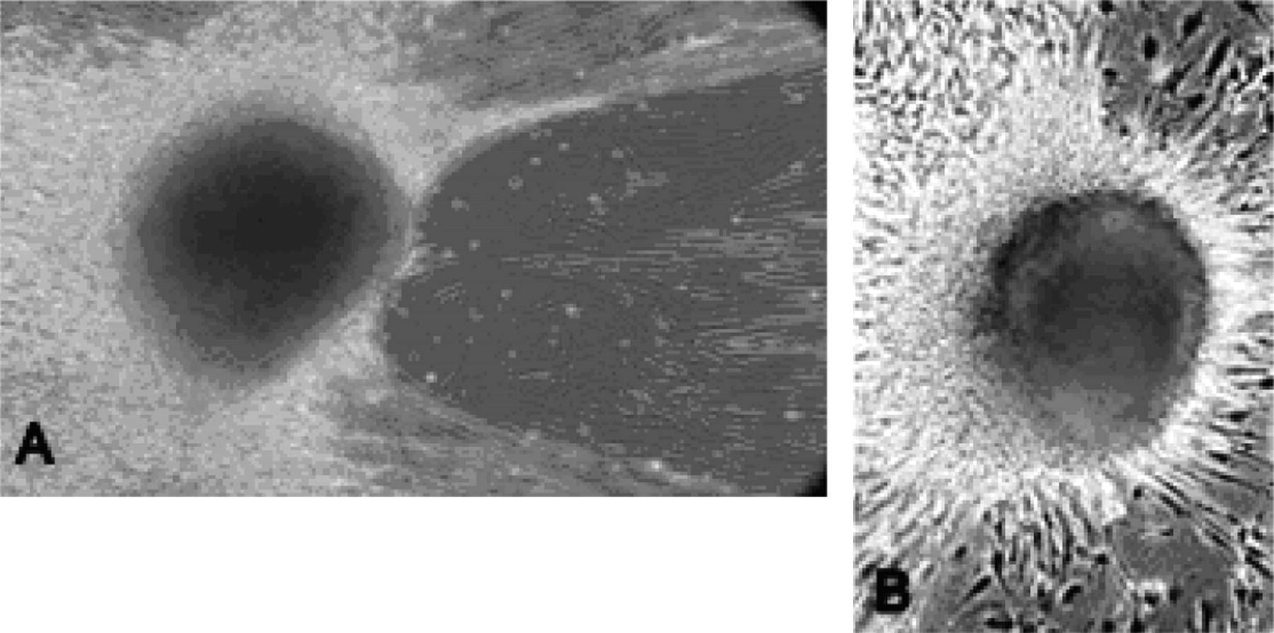
The CPE observed was not the disrupted, lysed, or fused monolayers that are most frequently associated with virus contamination. Rather, the CPE observed in the OOS results presented as large colonies of cells mounded off monolayers, in wells seeded with the MRC-5 indicator cell line and inoculated with the test article. These colonies were frequently macroscopic, exceeding 100 μm in diameter. The colonies typically had a bilateral symmetry (either rounded or oblong) and were clearly distinct from cell debris that was characterized by an irregular shape and not attached to the tissue culture plate. These structures resembled foci observed in tissue culture cells infected with transforming viruses. Figure 2 illustrates the similarities between the “foci” observed in the AVA and those seen in NIH 3T3 cells transformed with murine polyomavirus. An area of detached or disrupted cells was frequently observed along one side of the foci as seen in Figure 2A. When hemadsorption was noted, the erythrocytes were usually observed around the periphery of the disrupted area.

CPE observed in AVA OOS results resembled foci in Polyomavirus-transformed cells. A) Focus observed in test article-inoculated MRC-5 well from AVA. B) Focus observed in NIH 3T3 cells transformed with murine polyomavirus.
Initial Steps in Investigation into the OOS Results
One of the first steps taken in the investigation was to determine whether or not the OOS results could be reproduced. OOS results were obtained in repeat testing of sample retains. In addition, duplicate bioreactor samples yielded comparable OOS results. Bioreactor samples from different culture days also tested OOS with the exception of a single final harvest sample from a bioreactor for which other sampling days tested OOS.
Because the OOS results were obtained in testing of samples for a commercial product and development product manufactured at one site and the same development product campaigned at a second manufacturing site, the investigation assessed common factors between the sites and the products. The Working Cell Banks (WCB) for both products were exonerated, both through prior use in manufacturing multiple lots and because it was unlikely that WCBs manufactured at widely different times to support two unrelated products would have been contaminated.
Another common feature between products and sites was the testing lab used for the AVA. At the time of the apparent virus contamination, Centocor was using a contract lab, Contract Lab A, for the AVA of both products; so Centocor visited this lab. Although some areas for improvement in aseptic technique and training, as well as some potential points of sample cross-contamination, were noted, Centocor found no reason to invalidate the tests yielding OOS results or to suspect an assay artifact. Further, both Centocor and Contract Lab A conducted independent Kepner-Tregoe (K-T) analyses, using a structured, rigorous approach to attempt to identify the cause for the OOS results. Both Centocor and Contract Lab A predicated their K-T analyses on the assumption that the OOS results were generated in a valid test and concluded that these results were unlikely due to a contaminant originating at the testing lab.
OOS Results Attributed to Biological Agent Most Likely Introduced through Contaminated Medium
A number of initial observations suggested that the OOS results were caused by a biological agent. The results were reproducible, as previously discussed. In addition, the phenomenon was subcultivable, although no increase in apparent titer was observed following subcultivation. Further, the phenomenon was dilutable. The initial dilution series was only carried out to 1:100; no endpoint was determined and no decrease in apparent titer was observed. The OOS results obtained in the AVA and during the course of the investigation were considered weak results (i.e., one or a few foci were observed in a few wells of the six-well plate format). This suggested that the titer of the putative agent was at the limit of detection in the AVA assay. Thus, the lack of increase or decrease in titer observed following subcultivation and dilution, respectively, was attributed to the titer in the test articles being at the limit of detection of the assay.
Because the putative biological agent appeared at approximately the same time in a development product campaigned at two sites in two countries, medium or media components were considered the most likely point of introduction for a biological contaminant. The existence of open processing steps during the milling of the powdered medium strengthened this hypothesis. OOS results were also obtained in testing of samples for a commercial product manufactured using a different medium, but sharing several media components. However, initially only samples from one lot of this commercial product tested OOS among multiple lots evaluated at the same time. This result was very weak, with only one focus observed in single well of the six-well plate. Thus, this result was initially thought most likely due to cross-contamination at the manufacturing site or cross-contamination of samples at the contract lab (e.g., in biological safety cabinets, incubators, etc.).
Broad, All-Inclusive Analytical Investigational Plan to Identify the Putative Contaminant
The resemblance of the CPE observed in the OOS results to foci associated with transforming virus infections caused a great deal of concern, especially since the foci were observed on the cultures of the human cell line included in the AVA, MRC-5. Due to the appearance of these foci, murine, bovine, or human polyomavirus contamination was suspected, and testing for these viruses was heavily represented in the analytical investigation. However, evaluation was not restricted to these viruses or even to viruses. Rather, an all-inclusive approach to screen broadly for both viral and microbial agents was developed. All reasonable known technologies were utilized, and multiple, redundant, and orthogonal methods were deployed to avoid reliance on the sensitivity of any given method or susceptibility to limitations of any specific approach. The test articles included retains and duplicates of bioreactor samples that previously tested OOS and samples derived directly from AVAs (cell, supernatants, or both) where OOS results had been obtained. An overview of the analytical investigational plan is provided in Figure 3.

Analytical investigation plan overview: all-inclusive approach.
Negative Results Obtained in Extensive Testing for Polyomavirus
A number of polymerase chain reaction (PCR)-based methodologies were employed to screen for polyomavirus of rodent, bovine, and human origin. As shown in Figure 4, PCR evaluations targeting both T- and t-antigen, as well as VP-1, were utilized. In addition, the testing performed was sensitive for detection of all known strains of human polyomavirus (BK, JC, KI, WU, and Merkel). A faint band was observed in a pan-polyomavirus PCR. However in duplicate testing of a retain, the band was not observed for the test article, but was seen twice in a negative control sample. The sample yielding this result was derived from AVA test wells, but a negative result was obtained for the bioreactor sample evaluated in the AVA from which these test wells were generated. Thus, the band was attributed to nucleic acid present in the AVA growth media that contained fetal bovine serum (FBS). Another potential non-negative result was observed in a degenerate polyomavirus PCR where a faint band was observed for bovine polyomavirus. However, the product was not amplifiable and a specific TaqMan PCR of a retain was negative for bovine polyomavirus. In summary, the results for the extensive polyomavirus testing were negative.

Polyomavirus PCR screening results.
Positive Result for SV40 Attributed to Expression Vector Sequence
A positive result was obtained in PCR testing for another papovavirus, SV40. An initial Basic Local Alignment Search Tool (BLAST) search showed that no SV40 sequences were used in the expression construct of the production cell line. Although transformation of MRC-5 is very rare, there is a published report of SV40-transformation of MRC-5 (1). Thus, it initially appeared that a contaminating virus had been identified. However, a repeated BLAST search revealed the presence of the 5′ end of SV40 T-antigen in the expression vector. Thus, the “positive” result was a true result, but it represented an artifact of the genetic construction, not contamination of production cell cultures by an infectious papovavirus.
Nucleic Acid–Based Screening Assays for Additional Rodent and Bovine Viruses
A number of additional nucleic acid–based screening assays were employed in an attempt to identify a contaminant. These included the information technology (IT) version of the traditional serological mouse antibody production (MAP) screen, MAP-IT, which screens for the viruses that are typically present in the MAP panel, plus Mycoplasma, sp. The traditional serological MAP for 16 different mouse viruses was also performed. A PCR screening for seven different bovine viruses was conducted, as well as a rodent virus PCR covering 27 different mouse, rat, and hamster viruses. Negative results were obtained for all of these tests.
Screening for Potential Contaminants of Human Origin
Centocor consulted with Dr. Larry Anderson at the Centers for Disease Control to determine whether there were any viruses that might originate in humans and contaminate and transform MRC-5 cells. As per Dr. Anderson's suggestion, Centocor worked with Dr. Sue Tong to arrange a pan-adenovirus PCR, a human bocavirus PCR, and the pan polyomavirus PCR previously described. All of these screens produced negative results.
Non-Specific, Broad Screening Tests
In addition to testing for specific contaminants, the analytical plan also included non-specific, broad screening tests. A single potential virus-like particle was observed in transmission electron microscopy (TEM) of the test article from AVA test wells on the MRC-5 indicator cell line, but additional potential virus-like particles were not observed in extended analyses. A retrovirus-like particle was also observed in TEM of the supernatant sample from the myeloma indicator cell line, which is an expected result. An in vivo assay for inapparent viruses was conducted using suckling and adult mice, guinea pigs, and embryonated eggs, which provided no evidence of viral infection.
The broad screening undertaken also included highly sensitive, state-of-the-art methodologies, Triangulation Identification for Genetic Evaluation of Risks (TIGER) at Ibis Biosciences (Carlsbad, CA) and High-Throughput Massively Parallel Sequencing (MPS) at BioReliance Corporation (Glasgow, UK), to comprehensively screen for a wide variety of viral and microbial contaminants. The TIGER analysis uses mass spectrometry and base composition analysis following DNA amplification to screen for a very broad panel of virus families and genera, and also includes a pan-bacterial and pan-fungal screen. No viruses or other biological agents were detected by TIGER in bioreactor samples or in wells generated in the AVA. MPS is capable of detecting productively replicating viruses, as well as latent or transforming viruses, and compares contaminant-specific sequences to published databases in order to identify the contaminant. Very rare sequence hits were obtained for bovine viral diarrhea virus (BVDV) and other pestiviruses, but these were ultimately attributed to cellular sequences fused to BVDV genomes and misidentified in the GenBank identifications. The lack of BVDV infection was also supported by the inability of BVDV to infect human cells, including MRC-5, or rodent cells such as the mouse myeloma production cell line (2). Furthermore, no bovine material was used in the production bioreactor from which the sample was generated. A hit also was obtained for Dengue virus. Although Dengue virus can infect MRC-5 cells, it is known to be lytic in Vero cells. Vero cells were included in the AVA, but no cell lysis of Vero was observed in any of the OOS results. Further, there is no non-human reservoir known for this arbovirus. Thus, the Dengue virus hit was interpreted as an artifact due to sequence homology with BVDV. A hit for Torque Teno Virus was determined to be incorrectly annotated in GenBank and ultimately attributed to sequence for the scarab beetle, Trochalina. Hits for baboon cytomegalovirus (CMV) and human papilloma virus (HPV) were attributed to Alu SINES (short intervening nuclear sequences), noncoding “junk” DNA. There were hits for non-viral sequences including Cryptophya, algae, and Clitophilus, an edible mushroom, and Hanusia phi, a freshwater protozoan, but none of these represented an adventitious infection. In summary, the MPS analysis yielded no evidence for adventitious viral or microbial contamination. The non-viral hits were attributed to minor DNA contaminants in the media, water, or reagents used for the MPS analysis. These were not unexpected results and point to the power and sensitivity of this technique.
Extended Laboratory Investigation
As the negative analytical test results accumulated, the laboratory investigation was extended to include attempts to reproduce the OOS results at two alternate contract testing labs performing a comparable AVA. In the additional testing, the AVA was extended to 28 days and additional indicator cell lines (NIH 3T3, 324K, CHO, and MEF) were added for evaluation of some of the samples. Multiple samples that had tested positive at Contract Lab A tested negative at the other contract labs, even when the incubation period was extended and additional indicator cells were added.
The extended investigation also included evaluation of medium and pre-culture samples. Note that if virus were present in medium, it is highly unlikely that it would be present at a sufficient titer to be detected in a 3 mL sample, which is the volume evaluated in the AVA. Further, one of the products was manufactured using a chemically-defined media (CDM). Although there were some open processing steps in the preparation of the CDM dried powder that might serve as entry points for a viral contaminant from a human operator, it would be necessary to test in excess of one liter of reconstituted medium (20 g/L) to detect a single virion present assuming contamination of a 250 kg batch with 104 virions potentially present in an average sneeze. Thus, positive results from testing the CDM were extremely unlikely. Nonetheless, Contract Lab A reported positive results for medium samples. However, four pre-cultures tested negative in the AVA at Contract Lab A after being fed with the same medium batch that had previously tested positive. These were inconsistent and implausible results suggested artifacts in the test system.
Comparison of AVA Parameters in Attempt To Induce Formation of the Foci Artifact
Because the OOS results were obtained in the AVA at a single contract lab among three evaluated, methodological differences at the three different contract labs were investigated. One key methodological difference that Centocor believed might be associated with the formation of the foci artifact was the length of time the indicator cell monolayer is exposed to cell debris present in the test article. At Contract Lab A, the test article remains in the wells until the plates are fed at one week. In contrast, at Contract Labs B and C the test article is removed from the wells following one hour incubation or clarified by low-speed centrifugation prior to inoculation, respectively. As the test article is a lysate of cells in conditioned medium and can contain debris from up to 20 million cells per milliliter, it seemed plausible that the prolonged presence of cell debris on the MRC-5 monolayer in combination with another assay variable or variables might result in an abnormal growth pattern for the MRC-5, resulting in the formation of the foci artifact. FBS concentration in assay growth media was selected as a second assay parameter that varied between the contract sites (5% at Contract Lab A, 10% at Lab B, and 2% at Lab C) and would be expected to affect growth of MRC-5 cells. Centocor collaborated with Contract Labs A, B, and C to conduct a study evaluating the impact of both prolonged exposure of the monolayer to cell debris and FBS concentration of growth media on the formation of the foci artifact while using the standard method at each laboratory for all other assay parameters. A bioreactor test sample previously testing OOS at Contract Lab A was used as the test article. Contract Labs B and C reported negative results under all of the test conditions (i.e., clarified or non-clarified using 2%, 5%, and 10% FBS). OOS results were observed only at Contract Lab A, but they did not consistently appear in a treatment group (e.g., foci were not observed in all unclarified samples). See Table I for a summary of the results. Although the results were not conclusive, the data suggested that the formation of atypical foci structures might be related to some interaction of serum concentration and the prolonged exposure of the MRC-5 monolayer to cell debris.
Evaluation of Methodological Differences on MRC-5 AVA results Reported by the Different Contract Labs
Further Evaluation of Biological Nature of Putative Contaminant
An experiment was designed to determine an endpoint dilution for the putative contaminant with the thinking that it should be possible to determine an endpoint dilution of a biological contaminant, regardless of the nature of the contaminant. A bioreactor sample that had previously tested OOS was diluted using 10-fold dilutions to 10−7, and the dilutions were evaluated in the AVA. In this experiment, the undiluted sample that had tested OOS in prior testing tested negative. The 10−1 sample also tested negative, but the 10−2 dilution was OOS, showing foci. The rest of the dilution series tested negative. These results again were inconsistent and implausible, suggesting an artifact in the test system.
The effects of nanofiltration and gamma irradiation were evaluated for bioreactor and medium samples that had previously tested OOS, as both of these treatments would be expected to clear or at least reduce titer of most biological contaminants. Negative results were obtained for untreated samples that had previously tested OOS. Both the bioreactor sample and the medium sample tested negative following nanofiltration. However, after the samples were irradiated using 30 kGy, an OOS result was obtained for the bioreactor sample while the medium sample remained negative. These results were inconsistent because samples previously testing OOS tested negative, and they were implausible because a sample testing negative prior to treatment tested OOS following irradiation. These results are summarized in Table II.
Effect of Irradiation and Nanofiltration on Reproducibility of OOS Results
On-Site Evaluation of AVA at Three Contract Labs
An on-site evaluation of the AVA during actual laboratory execution was conducted at the three contract labs and included an exhaustive review of laboratory notebooks. At Contract Lab A, there were multiple laboratory notebook notations of irregularities in the negative control cultures that started at the time the OOS results were first reported. In addition, a Centocor staff member observed foci in both the test article-inoculated and the negative control samples for the MRC-5 indicator cell line. When questioned, the Contract Lab A staff indicated that these were consistent and typical results. However these results were never recorded in the test report. Figure 5A illustrates a structure observed in the negative control that was considered normal by Contract Lab A staff, while Figure 5B shows a structure that was considered an atypical OOS result. Contract Lab A staff indicated that a comparison of the size and shape of structures within any given assay allowed an experienced analyst to judge whether the result was negative or OOS. Foci were never observed at the other contract labs.

Comparison of structures observed in AVA during on-site review at Contract Lab A. A) Structure observed in negative control culture that was considered typical. B) Structure observed in test article–inoculated culture that was scored as atypical (OOS).
Identification of Potential Root Cause for Foci Artifact
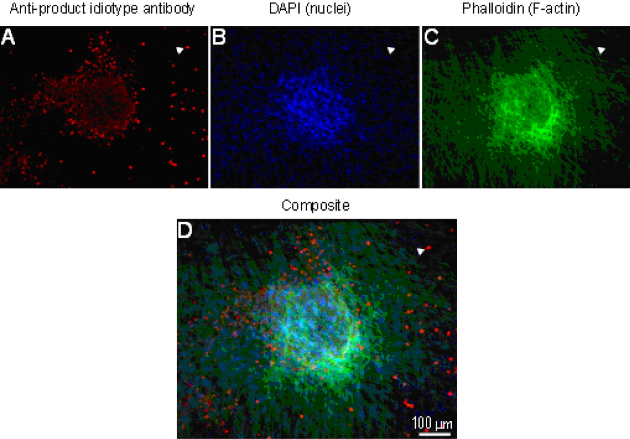
The inter-laboratory study suggested that the foci artifact might be related to an interaction of FBS concentration in the assay growth medium and the prolonged exposure of the MRC-5 monolayer to cell debris present in the test article. Centocor hypothesized that a clump of cell debris might serve a nidus for the formation of a focus and conducted immunofluorescence (IF) and TEM evaluations to characterize the cellular content of the foci in an attempt to discern a root cause for generation of the artifact. The foci examined in these characterization studies were generated at Contract Lab A.
For the IF experiments, cells were fixed and anti-product idiotype antibody was used in conjunction with an Alexa-488-conjugated secondary to detect test article cell debris. 4′,6-diamidino-2-phenylindole (DAPI) and Alexa-647 phalloidin were used to counterstain nuclei and filamentous actin, respectively. The stained foci were examined using confocal microscopy.
The IF study revealed that the foci were composed of one or more layers of intact cells surrounding a core of production cell line debris. In the composite image shown in Figure 6D, the blue-green color of the outer surface of the foci demonstrates convergence of blue stain for nuclei (Figure 6B) and green stain for actin (Figure 6C), suggesting that the outer surface of the foci consists of intact, viable MRC-5 cells. In contrast, the diffuse red staining for product observed in Figure 6A and the absence of this diffuse red signal in the composite image (Figure 6D) suggest that the core of the foci are composed of production cell debris from the test article and that this debris is covered by MRC-5 cells and thus is not present on the surface of the foci. The staining for product at the periphery of the core was more intense than that observed deeper within the core (Figure 6A). This staining pattern was more obvious with larger foci and suggests more pronounced deterioration of antigen within the foci. In addition to staining the foci, the product anti-idiotype antibody also stained cell debris that was distributed across the surface of the MRC-5 monolayer (Figure 6A and 6D). Although the staining pattern of this distributed debris suggested intact single cells, these “cells” were not counterstained with DAPI (compare signal at arrowheads in Figure 6A, 6B, and 6D), demonstrating that they did not contain nuclei and thus were not intact viable cells, but cell debris. The product staining pattern observed within the foci was diffuse compared to that observed with the control myeloma cells expressing product (data not shown) and for the cell debris on the MRC-5 monolayer. This diffuse staining pattern suggests cellular degradation, consistent with the absence of phalloidin staining of the foci core.

Immunofluorescence of AVA test well-generated as OOS result during routine testing at Contract Lab A. A. Staining pattern observed following incubation with rabbit anti-product idiotype antibody followed by an Alexa-488-conjugated donkey anti-rabbit IgG secondary antibody. B. Staining pattern observed following incubation with DAPI. C. Staining pattern observed following incubation with Alexa-647 phalloidin. D. Composite image showing co-localization of product, DAPI, and phalloidin signals.
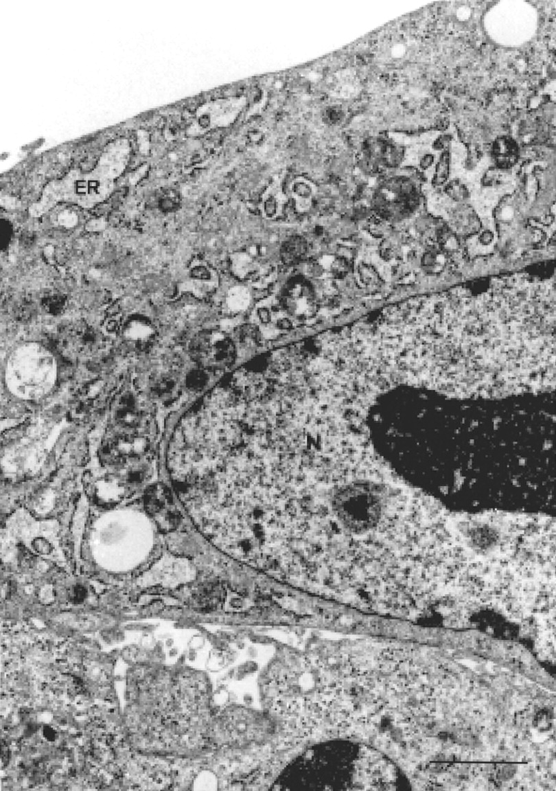
For TEM, foci plucked from the MRC-5 monolayers were fixed and dehydrated. Then thin sections were prepared and stained. The TEM analysis showed that the foci consisted of an outer periphery of intact cells surrounding an inner core of diffuse cell debris. The periphery consisted of varying numbers of cells showing the typical morphology and size of MRC-5 cells (Figure 7). In contrast, intact cells were not observed in the inner core of the foci. This core contained only cell debris (vesicles, membrane residues) (Figure 8). Although some electron-dense particles could be detected within the foci, no retrovirus-like particles (RVLP) were detected. Although presence of RVLP is characteristic of myeloma cells, RVLP are not stable (e.g., they do not typically maintain their distinct characteristic morphology when isolated from bioreactor culture supernatants). Thus, the absence of discernable RVLP in the cell debris is not instructive regarding cellular origin.

Ultrathin section through the periphery of focus. ER = endoplasmic reticulum, N = nucleus (magnification 10500×; bar = 2 μm).

Ultrathin section through the inner part of a focus (overview). CD: cell debris; (magnification 12500 ×; bar = 2.0 μm).
The results of the IF and TEM analyses indicated that the foci observed in test article-inoculated AVA wells were composed of a core of degraded myeloma cell debris covered by one or more layers of MRC-5 cells. The observation that the foci were detected only in the AVA at Contract Lab A where the MRC-5 monolayer is exposed to production cell line debris present in the test article for a prolonged period strongly suggests that these foci form when MRC-5 grow over myeloma production cell line debris present in the test article.
Overall Conclusion, Outcomes, and Lessons Learned
Centocor concluded that the OOS results were artifacts of the AVA test system at Contract Lab A and were not a result of contamination with a virus or other biological agent. Testing was discontinued at Contract Lab A due to the high risk of false positive test results. In addition, Centocor felt that remediation and requalification of the assay, as well as improvement of Contract Lab A's Quality Systems, were required. The AVA was validated at a Contract Lab B for the development project (the AVA at Contract Lab B was already qualified for the commercial product). Manufacturing of the commercial product resumed in consultation with health authorities and the marketing application for the late phase development product was approved. Impacted lots, as well as lots that had never tested OOS, were retested at Contract Lab B as per a Centocor OOS SOP and released.
Several factors supported this favorable outcome. Centocor acted quickly to quarantine lots and shutdown manufacturing, and then implemented a mitigation and decontamination plan that included H202 vapor decontamination of high-risk areas. Importantly, the company engaged in rapid, frequent, and open communications with the health authorities. As detailed in this article, extensive screening, including state-of-the art technology and a broad spectrum of sensitive methods, provided convincing evidence for the lack of an infectious viral or other biological agent. Finally, Centocor was able to follow an SOP that was already in place to retest impacted lots and ultimately disposition these for sale.
Several lessons learned were additional outcomes of this experience. Robust technical review of methods in use at contract testing labs is essential. If feasible, direct witness of laboratory execution and/or data assessment by a sponsor subject matter expert (SME) can be invaluable. A sponsor should thoroughly understand the method options that are allowed for in work instructions that may underlie the SOPs which the Sponsor normally reviews. Client protocols must be sufficiently detailed to eliminate undesirable method options. Audits should include a detailed review of laboratory notebooks; spot checks alone are not sufficient. An awareness of the training and experience of contract lab personnel must be maintained with appropriate risk mitigation considered for turnover of key personnel. Prearrangements should be made with contract labs for permanent documentation (e.g., photograph or fixed, stained assay plate) of atypical or OOS results in those assays where the results reported depend on the qualitative visual assessment of experienced personnel. Knowledge of all nucleic acid sequences present in expression vectors is required, including nonfunctional and/or partial sequences. This is especially important for interpretation of test results relying on nucleic acid sequence data. Sponsors should consider development of a Contamination Master Plan and designation of a single SME accountable for oversight of adventitious agent testing across all manufacturing sites. Sponsors should further consider treatment of medium as risk mitigation for virus and mycoplasma contamination. Although adaptation of animal component–free manufacturing processes reduces the risk of contamination with some potential viral and mycoplasma contaminants, it does not eliminate risk, as contaminants may be introduced by rodent or insect adulteration of warehoused medium or medium components or during open steps in preparation of dried medium powders.
- © PDA, Inc. 2010


Jump to section
- Article
- Abstract
- Introduction
- OOS Results Obtained in an In Vitro Adventitious Agent Assay
- CPE Observed Resembled Foci Seen in Polyomavirus-Transformed Cells
- Initial Steps in Investigation into the OOS Results
- OOS Results Attributed to Biological Agent Most Likely Introduced through Contaminated Medium
- Broad, All-Inclusive Analytical Investigational Plan to Identify the Putative Contaminant
- Negative Results Obtained in Extensive Testing for Polyomavirus
- Positive Result for SV40 Attributed to Expression Vector Sequence
- Nucleic Acid–Based Screening Assays for Additional Rodent and Bovine Viruses
- Screening for Potential Contaminants of Human Origin
- Non-Specific, Broad Screening Tests
- Extended Laboratory Investigation
- Comparison of AVA Parameters in Attempt To Induce Formation of the Foci Artifact
- Further Evaluation of Biological Nature of Putative Contaminant
- On-Site Evaluation of AVA at Three Contract Labs
- Identification of Potential Root Cause for Foci Artifact
- Overall Conclusion, Outcomes, and Lessons Learned
- References
- Figures & Data
- References
- Info & Metrics