
Abstract
A real-time polymerase chain reaction (RT-PCR) assay was developed to detect Burkholderia cepacia in pharmaceutical products contaminated with low levels of bacteria. Different pharmaceutical suspensions were artificially contaminated with B. cepacia, Escherichia coli, Staphylococcus aureus, and Bacillus megaterium. After a 24 h incubation in trypticase soy broth with Tween 20, samples were streaked on mannitol salt, phenyl ethyl alcohol, eosin methylene blue, MacConkey, and pseudomonas isolation agar. Microbial DNA was extracted from each sample by using a Tris-EDTA, proteinase K, Tween 20 buffer. Regular PCR targeting the 1.5 kilobases 16S rRNA eubacterial gene and cloning showed the predominant DNA in the extracted mix belonged to E. coli. Selective media isolation of bacterial contamination showed B. cepacia only detected on pseudomonas isolation while eosin methylene blue and MacConkey detected only E. coli. RT-PCR using primers PSL1 and PSR1 amplified a 209 bp 16S rRNA fragment using a Roche LightCycler 96® system with SYBR green I, a common double-stranded binding dye. The cycle at which fluorescence from amplification exceeds the background fluorescence was referred to as quantification cycle. All samples were found to be positive by standard microbiological testing and RT-PCR. B. cepacia was detected within 30 h in all contaminated samples using RT-PCR. Based upon standard curve analysis of B. cepacia DNA, the minimum DNA concentration that could be detected was 10 fg/uL with a correlation value of 0.98. RT-PCR detection of B. cepacia allowed faster quality control analysis, corrective actions, and process optimization.
LAY ABSTRACT: A real-time polymerase chain reaction (RT-PCR) assay was developed to detect Burkholderia cepacia in pharmaceutical products contaminated with low levels of bacteria. B. cepacia is the number one reason for microbial contamination recalls of non-sterile drug products in the USA. RT-PCR using primers PSL1 and PSR1 amplified a 209 bp 16S rRNA fragment using a Roche LightCycler 96® system with SYBR green I, a common double-stranded binding dye. All samples were found to be positive by standard microbiological testing and RT-PCR. B. cepacia was detected within 30 h in all contaminated samples using RT-PCR. RT-PCR detection of B. cepacia allowed faster quality control analysis, corrective actions, and process optimization.
Introduction
Microbial contamination of pharmaceutical products is still a threat to public health in the United States and around the world. Pathogenic microorganisms cause morbidity and in some cases mortality when present in pharmaceutical products (1). Furthermore, microbial breakdown of formulations can affect potency and shelf life. Based upon published scientific surveys, Burkholderia cepacia is the most frequently found microbial contaminant in sterile and non-sterile pharmaceutical products (1, 2).
Furthermore, a recent outbreak of B. cepacia complex (Bcc) pseudo bacteremia was associated to contaminated antiseptic formulations (3). During that outbreak, B. cepacia was isolated from blood cultures of 40 patients and antiseptic formulations. The outbreak investigation determined that the formulation was misused as a skin antiseptic during blood culture. The contaminated product was discarded and the staff retrained. Another outbreak was reported at a private hospital where 13 cancer patients undergoing chemotherapy developed B. cepacia bacteremia due to a contaminated antiemetic drug (4). The outbreak lasted 2 months and was controlled when hospital personnel were properly educated to optimize daily aseptic practices. Opened and unopened vials of the antiemetic drug grew B. cepacia. A recent contamination of a liquid pharmaceutical product triggered a multistate outbreak of B. cepacia infections in the United States (5). More than 53 cases were reported in five states. Furthermore, B. cepacia was also detected in the water system used for product manufacturing (6, 7).
The persistence of B. cepacia in pharmaceutical products can be explained by the lack of proper good manufacturing practice (GMP) and the use of compendial methods that do not provide the sensitivity and resolution to detect B. cepacia in pharmaceutical water, raw materials, and finished products (1, 2, 6). B. cepacia genetic and metabolic diversity are severely underestimated by industrial operators. The genome consists of more than one chromosome containing a wide variety of genes that are expressed to provide resistant to biocides, antibiotics, and adaptation to environmental stresses (8⇓⇓–11).
Most pharmaceutical companies relied on traditional cultivation and phenotypic methods to isolate and identify microbial contamination (1, 12). These methods are time consuming (5–7 days) and laborious. They relied upon the growth of microorganisms on the specific substrates in the media. However, some bacteria do not grow on those substrates or grow too slow to be detected by the incubation times currently used. In some cases, microbial cells undergo a physiological state by reducing metabolic reactions rates and cell size, which are affecting the protein and enzymatic profiles used to identify environmental isolates (1). These microbial cell changes are triggered by physical processes and environmental systems implemented to reduce or eliminate microorganisms during pharmaceutical manufacturing. If the processes and systems are not validated or properly implemented, microorganisms contaminate products, raw materials, and equipment (1, 12).
Rapid polymerase chain reaction (PCR) detection of B. cepacia contamination in pharmaceutical samples has been previously reported (13⇓–15). Studies showed PCR assays capable of enhancing detection and identification of B. cepacia, resulting in faster corrective actions and process optimization. Detection of amplified fragments was performed using gel electrophoresis. However, there are no reported scientific studies on the application of real-time PCR (RT-PCR) to detect B. cepacia contamination in pharmaceutical samples. Nor has any RT-PCR assay been developed for detecting B. cepacia in formulations such as liquid, tablets, and cream contaminated with low numbers of bacteria. The objective of this study was to develop a RT-PCR protocol for the detection of B. cepacia in different pharmaceutical delivery systems such as tablets, creams, and liquid formulations contaminated with mixed bacterial cultures.
Materials and Methods
Samples
Different types of pharmaceutical formulations were analyzed. They ranged from over the counter liquid, creams, and tablet formulations. The products were purchased at the local CVS pharmacy store located in Fair Lawn, NJ, USA.
Microorganisms
The microorganisms spiked into product suspensions were: B. cepacia ATCC 25416, Staphylococcus aureus Ward Science (wardsci.com), Bacillus megaterium Ward Science (wardsci.com), and Escherichia coli Ward Science (wardsci.com). B. cepacia ATCC 25416 is the type strain for genomovar I of the Bcc. The bacterial species used in this study were commonly found in pharmaceutical products, raw materials, and water systems (1, 7). All microbial identifications were confirmed by16S rRNA sequencing.
Inoculum Preparation
Bacterial cultures of E. coli, B. megaterium, and S. aureus grown in trypticase soy broth (TSB) Ward Science (wardsci.com) were shaken, 200 rpm, for 24 h at 37 °C. B. cepacia cultures were shaken for 48 h. After incubation, all bacteria were diluted with deionized sterile water to obtain a final inoculum ranging from 10 to 100 CFU/mL.
Sample Inoculation and Incubation
One gram of each pharmaceutical product was added to 100 mL of TSB containing 2% or 4% Tween 20. After product addition, samples were inoculated with serial dilutions of all four bacteria followed by incubation at 37 °C for 24 h with shaking at 200 rpm.
Microbiological Analysis of Samples
After incubation in TSB, samples were streaked onto the following selective agar media: pseudomonas isolation (PIA), MacConkey (MAC), eosin methylene blue (EMB), phenyl ethyl alcohol (PEA), and mannitol salt (MSA). All selective media plates were incubated for 48 h at 37 °C. Random colonies, based upon colony morphology and color, grown on selective media plates were identified by Gram stain, biochemical, and genetic analysis.
DNA Extraction
Bacterial DNA was extracted from contaminated product suspensions and colonies using a Tris-EDTA buffer containing Tween 20 and proteinase K. Extractions were incubated for 20 min at 37 °C followed by 10 min at 95 °C. Different aliquots of extracted bacterial DNA were used in the PCR reactions.
B. cepacia DNA was extracted from pure cultures as described in the ZR Soil Microbe DNA MiniPrep protocol (Zymo Research, Irvine, CA). Fluorometric quantitation of bacterial DNA concentration was determined using the Qubit® dsDNA assay procedure (Thermo Fisher Scientific, Waltham, MA). After quantitation, ten-fold serial dilutions of B. cepacia DNA were used as standard curves. Negative controls based upon sterile water were included in RT and regular PCR assays. All experiments were performed in duplicate.
Standard PCR
Bacterial DNA extracted from contaminated product suspensions was amplified using PCR beads as previously described (16). The amplification reactions targeted the 1.5 kilobases (kb) 16S rRNA eubacterial gene. Amplicon detection was carried out by gel electrophoresis using the FlashGelTM system (Lonza Inc., Rockland, ME) with FlashGel DNA Cassettes containing 1.2% agarose. A FlashGel DNA Marker (Lonza Inc., Rockland, ME) with fragment sizes ranging from 100 bp to 4 kb was used to determine the presence of correct DNA gene fragments.
Cloning Libraries of Bacterial 16S rRNA Genes
The DNA fragments from the PCR amplification of eubacteria 16S rRNA genes were cloned using plasmid pCR®4-TOPO (Life Technologies, Thermo Fisher Scientific, Grand Island, NY) according to the manufacturer's instructions. Transformations were performed using Mix and Go Competent E. coli strains (Zymo Research, Irvine, CA). White colonies grown on Luria-Bertani (LB) agar with ampicillin (50 ug/mL) were transferred to LB broth containing ampicillin (50 ug/mL). Samples were incubated overnight at 37 °C.
Plasmids were isolated from each clone using the Zyppy Plasmid Miniprep Kit (Zymo Research, Irvine, CA). Cloned inserts were reamplified using M13 DNA primers. DNA sequencing reactions of the amplified PCR fragments were performed by a contract-testing laboratory (Genewiz Inc., South Plainfield, NJ). Homology searches for 16S rRNA genes were performed using the Classifier application on the Ribosomal Database Project (http://rdp.cme.msu.edu/classifier/classifier.jsp) (17) and also using the GenBank server of the National Center for Biotechnology Information (NCBI) (http://blast.ncbi.nlm.nih.gov/Blast.cgi) and the BLAST algorithm (18).
RT-PCR Reactions
Bacterial DNA extracted from artificially contaminated product suspensions was used to develop the RT-PCR assay. RT-PCR was performed with a LightCycler® 96 System (Roche Life Science, Indianapolis, IN) using SYBR green PCR master mix as the detection system in a reaction mixture of 25 uL. Aliquots of 3 uL of extracted bacterial DNA from pure cultures and product suspensions were used for all reactions. DNA primers PSL1 and PSR1 amplified a 209-base pair fragment of the B. cepacia 16S rRNA gene (14, 19). Different reaction conditions and cycles were analyzed to optimize target detection. However, the optimized reaction conditions were a preincubation cycle of 95 °C for 600 s, three-step amplification of 30 cycles of 95 °C/10 s, 58 °C/10 s, 72 °C/20 s, and a melting cycle of 95 °C/10 s, 65 °C/60 s, 97 °C/15 s. Gel electrophoresis analysis was performed to confirm the presence of the 209 bp fragment. The cycle at which fluorescence from amplification exceeds the background fluorescence was referred as quantification cycle (Cq). B. cepacia DNA concentration in artificially contaminated drug products was determined using a standard DNA curve as described above. A standard curve displays a graph of Cq values against the base 10 logarithm of the quantity of each standard. For absolute quantification, the absolute values of the standard curve were used to assign quantities to unknown samples.
R^2, the coefficient of determination is the proportion of variability in a data set that is accounted for by a statistical model. The intercept is the expected mean value of y when all x = 0. The slope of a regression line (b) represents the rate of change in y as x changes. Error is the deviation of the observed value from the true value of a quantity of interest. Efficiency is the measure of quality of an estimator, of an experimental design, or a hypothesis testing procedure. The Cq and standard curve analysis were performed using the LightCycler® 96 System Instrument software version 1.1.
Results and Discussion
Conventional methods relying on selective agar media after enrichment in TSB containing 2% Tween 20 were ascertained to detect B. cepacia in pharmaceutical samples with low levels of a mixed bacterial contamination. Pharmaceutical dosage forms such as tablets, creams, and liquid formulations were artificially contaminated with a mixed bacterial culture containing B. cepacia, E. coli, S. aureus, and B. megaterium. The selected bacteria were commonly found or are representatives of the types of bacteria found in products recalled in the USA (1, 2). Bacterial cultures were spiked into each drug product suspension with colony-forming unit (CFU) numbers below 75 CFU/mL. The average CFU bacterial inoculum for B. cepacia and E. coli were 14 and 62 CFU/mL, respectively. While B. megaterium and S. aureus showed average inoculum counts of 3 and 42 CFU/mL, respectively.
After inoculation, artificially contaminated drug samples were shaken for 18–24 hours at 37 °C to promote bacterial growth. Table I shows the results for each selective agar media analyzed. Out of seven products tested, only three products showed bacterial growth on all plates. These samples were itch stopping cream, calamine anti-inflammation and pain lotion, and liquid insect repellent. Identification of colonies growing on selective agar media for gram-positive bacteria, MSA and PEA, showed S. aureus while all EMB and MAC plates were found to have E. coli (Table I). PIA agar plates for those three samples demonstrated the presence of B. cepacia after a 48 h incubation at 37 °C. Four more products showed B. cepacia isolation on PIA media. These products were liquid antiseptic mouthwash, triclosan dentifrice, and cortisone anti-inflammatory cream. MAC has been previously used to recover B. cepacia from contaminated products (1, 2). However, if there are other gram-negative bacteria present such as E. coli, detection can be inhibited due to the high growth rates of E. coli and other gram-negative bacteria when compared to B. cepacia. The use of selective agar for B. cepacia such as B. cepacia selective agar (BCSA) can enhance the isolation and detection from mix culture contaminations (6). PIA was selective enough to recover B. cepacia from all products. However, to isolate B. cepacia on PIA from hydrocodone acetaminophen tablets, the Tween 20 concentration in TSB was increased from 2% to 4%. Lower concentrations than 4% did not recover B. cepacia from tablets. The 4% Tween 20 concentration neutralized the active ingredient in the hydrocodone acetaminophen tablets allowing the growth and detection of B. cepacia. The neutralization of the antimicrobial ingredients in the pharmaceutical formulations is a very important step in the isolation of B. cepacia. Unfortunately, this is commonly overlooked, resulting in false negative reactions when microbiology methods are not properly validated (1, 12).
Isolation of Bacterial Contamination on Selective Media and Identification of Predominant Colony Type from Pharmaceutical Products Contaminated with Mixed Bacterial Cultures
Contaminated pharmaceutical samples inoculated with the mixed culture were analyzed by a quick DNA extraction procedure using Tris-EDTA buffer, Tween 20, and proteinase K. The procedure lasted only 30 min and did not require organic solvents or hazardous chemicals. To determine the predominant bacterial DNA in the extracted product suspensions, a standard PCR reaction was performed targeting the 1.5 kb 16S rRNA eubacterial gene. The 1.5 kb fragment is a universal gene present in all members of the domain bacteria (16). The gene has conserved and variable regions that can be used to distinguish bacterial phyla, genera, and species. The amplified fragments from product suspensions were cloned into vector plasmid pCR®4-TOPO. DNA sequencing of the clones demonstrated the predominant presence of E. coli in the clone library. No other bacterial 16S rRNA sequence was detected. Evidently, E. coli outcompeted B. megaterium, S. aureus, and B. cepacia in all contaminated samples. This was also confirmed with bacteria identified on MAC and EMB selective media plates, which were shown to be E. coli.
To specifically detect B. cepacia in the extracted bacterial DNA, a 209 bp fragment of the B. cepacia 16S rRNA gene was used as a target to develop a RT-PCR assay. The copy number for the 16S rRNA gene is 6 (8, 19). The genome size for B. cepacia strain ATCC 25416 is 8.1 kb with four replicons of 3.65 megabases (MB), 3.17 MB, 1.07 MB, and 200 kb in size (8). The largest replicon has four copies of the ribosomal gene while the other two MB replicons have one each.
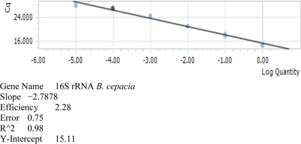
The sensitivity and accuracy of the primer pair PSL-1 and PSR-2 were previously described (14, 19). Standard curves of B. cepacia DNA were constructed to determine the DNA concentration of B. cepacia in pharmaceutical samples contaminated with B. megaterium, E. coli, S. aureus, and B. cepacia. The DNA concentration for undiluted samples of B. cepacia DNA was found to have 9.96 × 10⁻¹ ng/uL with an average Cq of 14.56 (Table II). When ten-fold dilutions of B. cepacia DNA were tested, the minimum concentration detected was 9.96 × 10⁻⁶ ng/uL with a correlation coefficient of 0.98 (Figure 1). This concentration was equivalent to 10 femtograms (fg)/uL of B. cepacia DNA.
RT-PCR Detection of B. cepacia DNA Dilutions

Standard curve analysis of B. cepacia DNA dilutions after RT-PCR.
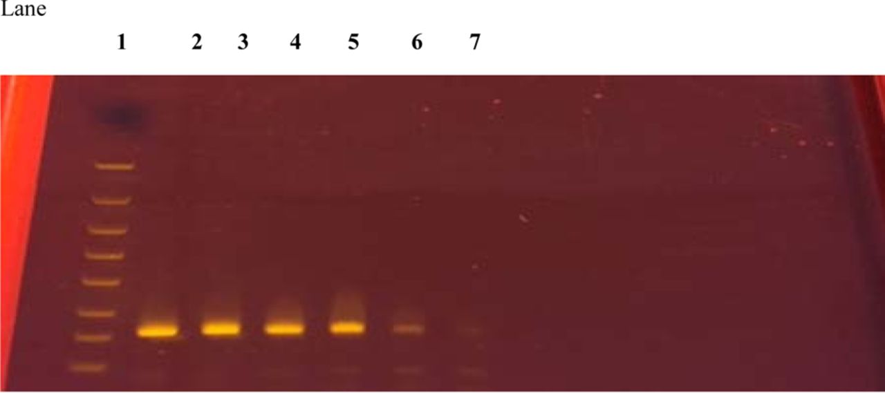
The Cq for the lowest DNA concentration detected was 27.99. Melting curve analysis of the amplified fragments confirmed the presence of the 209 bp fragment. Gel electrophoresis of the amplified fragments from standard curve samples was also positive, indicating the presence of the 209 bp fragment (Figure 2).

Gel electrophoresis of RT-PCR results for standard dilutions of B. cepacia DNA.
1 = Marker
2 = [DNA] = 9.96 × 10⁻¹ ng/uL
3 = [DNA] = 9.96 × 10⁻² ng/uL
4 = [DNA] = 9.96 × 10⁻³ ng/uL
5 = [DNA] = 9.96 × 10⁻⁴ ng/uL
6 = [DNA] = 9.96 × 10⁻⁵ ng/uL
7 = [DNA] = 9.96 × 10⁻⁶ ng/uL
Bacterial DNA extracted from contaminated sample suspensions was used in the RT-PCR assay. Different aliquots were analyzed but the optimal detection was found to be with 3 uL of extracted sample in a 25 uL reaction volume. Sterile water, DNA primers, and SYBR green PCR master mix were added to complete each reaction. Preliminary studies were performed to determine the number of cycles needed to detect B. cepacia in products contaminated with mixed bacterial cultures. The optimal number of cycles to perform the RT-PCR reaction was found to be 30 with no false-positive or false-negative reactions detected.
All contaminated products analyzed were found to be positive by the RT-PCR assay. Table III shows the average Cq for each pharmaceutical sample tested. The Cq indicated the cycle at which fluorescence from amplification exceeded the background fluorescence. Negative controls did not show any reaction and were completely devoid of any signal or band as per melting curve and gel electrophoresis analyses. Cq values for artificially contaminated pharmaceutical products ranged from 18.85 to 26.74. The faster Cq values were found with triclosan dentifrice samples with an average of 18.85 while hydrocodone acetaminophen tablets exhibited the longest, with an average of 26.74. When compared to the standard curve, all contaminated products exhibited Cq values way above the minimum detected B. cepacia DNA concentration, which was 27.99. This indicated that the growth conditions and neutralization protocol provided optimal conditions for B. cepacia to grow enough in the presence of other bacteria so the DNA can be extracted from the bacterial mixed culture using a mild DNA extraction protocol and subsequently detected by the RT-PCR. Evidently, the low B. cepacia inoculum with an average of 14 CFU/mL was easily detected after a 24 h enrichment.
RT-PCR and PIA Detection of B. cepacia in Pharmaceutical Products Contaminated with Mixed Bacterial Cultures
Rapid PCR detection of B. cepacia in contaminated products optimized quality control analysis and can provide a rapid response to any major outbreak due to the use of contaminated material (13⇓–15). However, samples analyzed from recent outbreaks caused by contaminated liquid docusate sodium products were plated on selective agar media (BCSA), which required 16.5 days for colonies to be visible and identified (6). Furthermore, positive samples were confirmed by molecular typing by a different laboratory, which probably required a couple of more days for final identification. However, the product enrichment (18–24 h), DNA extraction (30 min), and the RT-PCR procedure (3 h) developed in this study was completed in less than 30 h. Microbial DNA extraction from product suspensions was completed in less than 35 min. The RT-PCR setup and the thermocycler run were completed within 3 h. Data analysis did not take more than 10 min. Because the assay is detecting the amplified product in real time, the instrument screen provides ongoing real-time data analysis where the amplification curves can be used to predict a presumptive positive result before the assay is completed.
Shaking samples overnight and optimal neutralization of antimicrobial ingredients from each pharmaceutical formulation allowed faster detection times and assay resolution. For instance, the RT-PCR assay detected B. cepacia in hydrocodone acetaminophen tablets using TSB with 2% Tween 20 concentration. The RT-PCR signal was strong and the gel electrophoresis results did show a 209 bp band. However, B. cepacia was not recovered on PIA. Similar results were reported in previous studies with contaminated pharmaceutical syrups where the PCR assay detected B. cepacia but no positive detection was found using agar media (15). In that type of situation, if the analyst decided that the absence of growth on selective media plates indicated the absence of B. cepacia from the sample, it would have led to a false-negative report. Selective agar recovery was necessary to show equivalency with compendial methods but optimal neutralization of the formulations antimicrobial ingredients was also needed (1, 11). Some pharmaceutical companies and hospitals use MAC or EMB instead of PIA or BCSA for the isolation of gram-negative bacteria (3). Using MAC or EMB might underestimate B. cepacia contamination when other gram-negative bacteria are present. In our study, B. cepacia was not isolated using MAC or EMB because it was outcompeted by E. coli. However, using specific 16S rRNA sequences the RT-PCR assay was capable of detecting B. cepacia in a mixed culture of gram-negative and gram-positive bacteria. As non-sterile drug products may have a bioburden that may outcompete B. cepacia, RT-PCR has an advantage over culture methods.
B. cepacia is not only a health threat when it comes to morbidity and mortality but also because the stability and efficacy of products are compromised by the degradation of active ingredients and excipients (3, 10, 13, 20). Unfortunately, even at the beginning of the twenty-first century and despite all the substantial historical data, B. cepacia genetic and metabolic diversity continue to be severely underestimated by industrial operators, resulting in the morbidity and mortality of susceptible populations (5). However, regulatory agencies in the USA are promoting the addition of B. cepacia as an objectionable microorganism to compendial chapters (12). This will provide further guidance to companies, creating more awareness of the seriousness and challenge to provide efficacious and safe pharmaceutical products.
In conclusion, a RT-PCR assay was developed and validated to detect B. cepacia in pharmaceutical products contaminated with low numbers of mixed bacterial cultures. All samples showing positive results with standard microbiological methods were also positive with the RT-PCR. Rapid assessment of pharmaceutical samples provides important information on the quality control and possible risk of contaminated material, allowing the expeditious implementation of corrective actions to prevent morbidity and mortality by the lack of process control during manufacturing of pharmaceutical products. The recent outbreaks of B. cepacia demonstrated the need to not only apply molecular analysis to the identification of isolates from selective media plates but also to screen samples by PCR or other molecular methods that will provide a better resolution and sensitivity than standard methods before releasing products to the market. We also agree with a current statement by Marquez et al. (6) where they recommended the addition of Bcc as an objectionable organism for any non-sterile medication based upon the product's intended purpose and delivery system. Future studies will determine the applicability of the RT-PCR assay to quantify B. cepacia in pure and mixed bacterial cultures without prior enrichment of product suspensions.
Conflict of Interest Declaration
The authors declare that they have no competing interests.
- © PDA, Inc. 2018
Reference


Jump to section
Related Articles
Cited By...
- Identification of Burkholderia cepacia Complex by PCR: A Simple Way
- Design, Development, and Validation of a Culture-Independent Nucleic Acid Diagnostics Method for the Rapid Detection and Quantification of the Burkholderia cepacia Complex in Water with an Equivalence to ISO/TS 12869:2019
- Burkholderia cepacia Complex Bacteria: a Feared Contamination Risk in Water-Based Pharmaceutical Products